Figure 2.

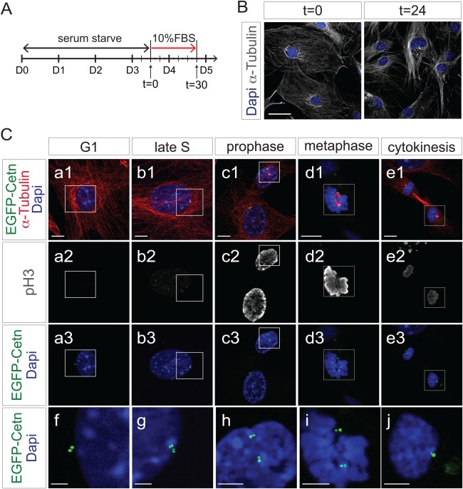
EGFP‐Centrin1 mirrors endogenous centrosome duplication cycle. A: Diagram of time‐course experiment shown in B and C. B: Fluorescence microscope images of ROSA‐EGFP‐Cetn1 MEFs in quiescence after serum starvation (t = 0), and in proliferation 24 h after serum induction (t = 24). Cells were immunostained with antibody to α‐Tubulin. Scale bar: 50 µm. C: Time‐course experiment following synchronization of ROSA‐EGFP‐Cetn1 MEFs. Cells were immunostained with antibodies to α‐Tubulin and Serine 10 phospho‐Histone H3 (pH3). (f, g, h, i, j) Magnified pictures of square area in of (a, b, c, d, e). Note centrosomes are duplicated by the end of S phase, and each centrosome is allocated into each dividing cell. Scale bars: 10 µm (a, b, c, d, e), 5µm (f, g, h, i, j).
