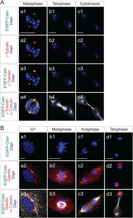
Figure 4.

Correlation of EGFP‐Centrin1 with γ‐Tubulin or Gogi apparatus in ROSA‐EGFP‐Cetn1‐MEFs. A: Fluorescence microscope images of ROSA‐EGFP‐Cetn1 MEFs in metaphase (a1‐4), telophase (b1‐4), or cytokinesis (c1‐4). Cells were immunostained with antibodies to γ‐Tubulin and α‐Tubulin. Note EGFP‐Centrin1 co‐localizes with γ‐Tubulin. Scale bars: 10 µm. B: Fluorescence microscope images of ROSA‐EGFP‐Cetn1 MEFs in G1 phase (a1‐3), metaphase (b1‐3), anaphase (c1‐2), or cytokinesis (d1‐3). Cells were immunostained with antibodies to Giantin and α‐Tubulin. Note Golgi apparatus localizes adjacent to EGFP‐Centrin1 although Golgi apparatus dispersed throughout the cytosol in metaphase and anaphase (b2 and c2). Scale bars: 10 µm.
