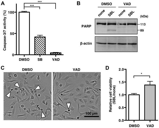
Figure 5.
SBL induces caspase-3/7 activation. (A) Caspase-3/7 activity in MDA-MB231 cells treated with or without 2 μM SBL in the presence of solvent DMSO, 10 μM p38 MAPK inhibitor SB203580 (SB) or 100 μM pan-caspase inhibitor zVAD-fmk (VAD) was assessed using SensoLyte Homogeneous AMC Caspase-3/7 assay kit. Each bar indicates the ratio of caspase-3/7 activity of the cells with SBL treatment to without SBL treatment. Results are means ± SD for two independent experiments conducted in triplicate. ***P<0.001. (B) Cell lysates of MDA-MB231 cells treated with or without 2 μM SBL in the presence of DMSO or 100 μM zVAD-fmk (VAD) for 72 h were subjected to immunoblotting to detect PARP and β-actin. The smaller size of PARP, approximately 89 kDa, is a cleaved form of PARP, which is correlated to caspase-3/7 activation. (C) Cell morphology of MDA-MB231 cells after treatment with SBL for 72 h in the presence of solvent DMSO or 100 μM zVAD-fmk (VAD). Arrowheads indicate dying or dead cells. (D) MDA-MB231 cells were treated with or without 2 μM SBL in the presence of DMSO or 100 μM zVAD-fmk (VAD) for 72 h. Each bar indicates the ratio of cell viability with or without SBL treatment. *P<0.05.

