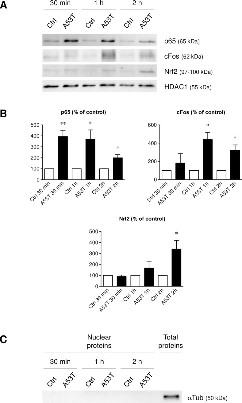
Fig 7. A53T protein induces NFkB, AP-1 and Nrf2 recruitment.
Nuclear localization of the p65 subunit, c-Fos (subunit of AP-1) as well as Nrf2 was evaluated on microglial cells after a 5 μM A53T exposure for 30 min, 1 h or 2 h (Fig 7A). These chemiluminescent detection assays were realized by western-blot with 10 μg of nuclear proteins. The histone deacetylase 1 (HDAC1) was used as a loading control. Furthermore, p65, c-Fos and Nrf2 detected proteins were normalized and quantified (Fig 7B). Results are given as mean ± SEM of at least three independent experiments. * p < 0.05, ** p < 0.01, significantly different from control condition. A cytoplasmic protein negative control western-blot (αTub) was also realized (Fig 7C) to demonstrate the effectiveness of the nuclear isolation.

