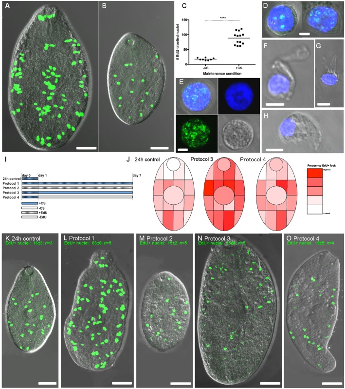
Fig 6. Proliferating cells in growing juvenile Fasciola hepatica.
Incorporation of 5-ethynyl-2-deoxyuridine (EdU) identifies DNA synthesis occurring during proliferation of cells with neoblast-like morphology. Green fluorescence denotes EdU and blue fluorescence denotes Hoechst 3342 labelling of nuclear DNA. A, B—Distribution of EdU labelled nuclei (EdU+) in fluke grown for 7 days in RPMI+50% chicken serum (A), or unsupplemented RPMI (B); C—Quantification of EdU+ nuclei in non-growing (-CS) vs growing (+CS) specimens; D, E—Morphology of dispersed EdU+ cells; D shows two example cells, E shows single cell and individual fluorescence signals (Hoechst 3342, EdU, brightfield, overlaid); F, G, H—Examples of non-proliferating (EdU-) cells showing distinct morphologies associated with differentiated cells; I, Pulse chase protocols; J, Heatmaps illustrating the change in EdU+ localisation associated with pulse-chase exposure, suggesting that EdU+ nuclei migrate towards differentiated tissue; K-O, example staining patterns of juvenile F. hepatica from each pulse-chase exposure protocol.

