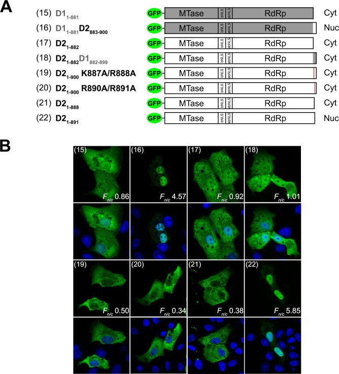
Fig 3. DENV1 and 2 NS5 C-terminal residues 883–900 confer differential sub-cellular localization.
(A) Schematic diagram of full-length and truncated DENV1 and 2, and chimeric full-length DENV1/2 fused to GFP. The plasmid annotation is as described in Fig 2A. (B) The GFP-NS5 protein constructs described in A were transfected into Vero cells and fixed at 24-hour post-transfection. Anti-GFP (ab6556 IgG, 1:1000) antibody was used for immunostaining and digitized images were captured by Zeiss LSM 710 upright confocal microscope by 40× oil immersion lens. The construct numbers used in Fig 3A are indicated in parenthesis in the images. Nuclear to cytoplasmic fluorescence ratio (F n/c) as previously described [17, 29, 30, 42] are indicated and data are shown as mean F n/c, n ≥ 30 cells from a single assay, representative of two independent experiments.

