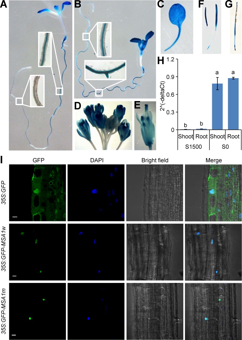
Fig 3. Expression pattern and subcellular localization of MSA1.
(A-G) Histochemical GUS staining of MSA1 promoter-GUS transgenic plants. One-week-old seedlings grown on agar solidified MGRL media with 1500 μM sulphate (A) or without sulphate (B). (C) A leaf from a two-week-old plant grown on agar solidified MGRL media with 1500μM sulphate; (D) the inflorescence of a plant grown in soil; (E) a flower; (F) developing siliques; (G) a mature silique. (H) Expression of MSA1 was strongly induced by S-deficiency. Plants were grown in S sufficient conditions (S1500) or S deficient conditions (S0) for two weeks. Expression level of MSA1 was normalized to the internal control gene UBQ10, and presented as 2^(-deltaCt) with means ± SD (n = 3). Columns with different letters indicate significant differences (P ≤ 0.01, LSD test). (I) Subcellular localization of MSA1. Constructs encoding GFP alone and GFP fused of wild type MSA1 (GFP-MSA1w) and mutated MSA1 (GFP-MSA1m) were transformed into Arabidopsis under the control of the CaMV 35S promoter. The GFP-MSA1 fusion protein was specifically expressed in the nucleus as stained by DAPI. Scale bar, 10μm.

