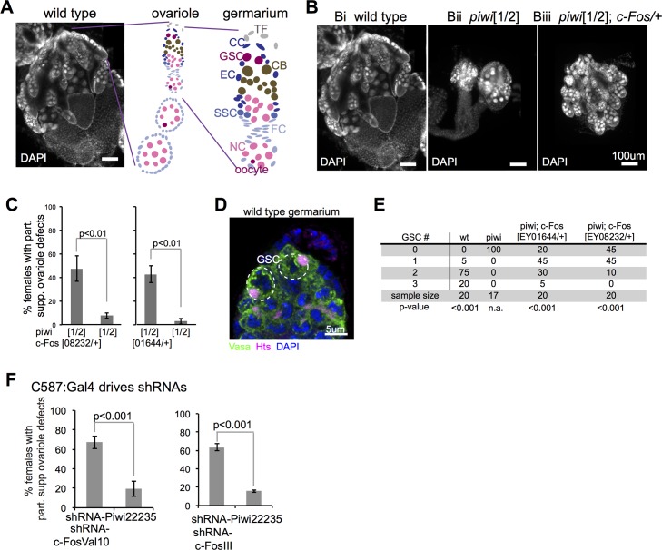
Fig 1. c-Fos reduction partially suppressed defects in piwi mutant ovaries.
(A) DAPI-stained image of a wild-type ovary, a diagramed ovariole, and a diagramed germarium. Labeled cell types are, respectively, TF, terminal filament; CC, cap cell; GSC, germline stem cell; CB, cystoblast; EC, escort cell; SSC, somatic stem cell; FC, follicle cell; NC, nurse cell; oocyte. (B) Images of DAPI-stained ovaries from wild-type and mutant animals. (C) Quantification of Drosophila females with large (partially suppressed ovariole defects as shown in 1Biii) ovaries in piwi[1/2] and c-Fos/+;piwi[1/2]. (D) Vasa (green) and Hts (magenta) IF and DAPI (blue) staining of a wild-type germarium, with white dashed circles around GSCs. (E) Quantification of GSCs per germarium. (F) Quantification of Drosophila females with large (partially suppressed ovariole defects as shown in 1Biii) ovaries in animals with C587:Gal4 (escort cell-specific) driving Piwi and c-Fos shRNAs. Error bars represent standard deviation, and the chi-square test was used for statistical comparison.

