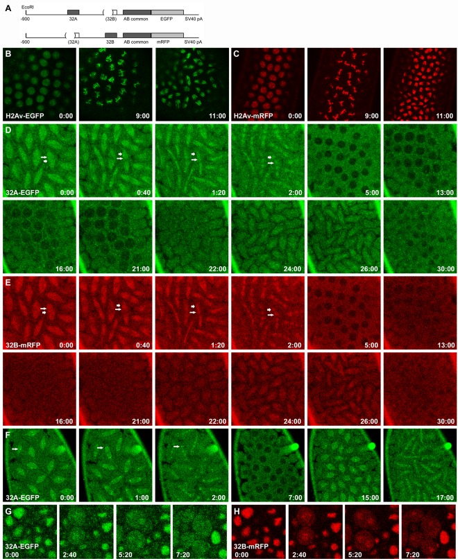
Fig 5. Dynamics of 32A and 32B during mitosis.
A. Schematic of the 32A-EGFP and 32B-mRFP transgenes. Genomic sequences are present from -900 to the codon for the last amino acid of BEAF, with the indicated sequences deleted so only 32A or 32B can be produced while allowing expression from endogenous BEAF sequences. B, C. Dynamics of H2Av-EGFP or H2Av-mRFP, respectively, during interphase and the metaphase and anaphase stages of mitosis in syncytial embryos. See also S1 and S2 Videos. D, E. Time course showing the dynamics of 32A-EGFP or 32B-mRFP, respectively, during two rounds of mitosis in the same syncytial embryo. Short, thick arrows point to chromosomal locations (metaphase at t = 0:00, anaphase to telophase in subsequent panels); longer, thin arrows point to what appears to be the mitotic spindle. See also S3 Video of this embryo and, for other examples, S4–S6 Videos. F. Time course showing the dynamics of 32A-EGFP during two rounds of mitosis in a syncytial embryo. Bright spots of 32A-EGFP that appear to be centromeric are prominent in this embryo (arrows). See also S3 Fig and S4–S6 Videos. G, H. Time course in an embryo during germ band elongation showing the dynamics of 32A-EGFP or 32B-mRFP, respectively, during mitosis. See also S7 Video. All times are in min:sec. Note that all embryos have a wild-type BEAF gene, but similar results were obtained in a BEAFAB-KO background. See text for details.

