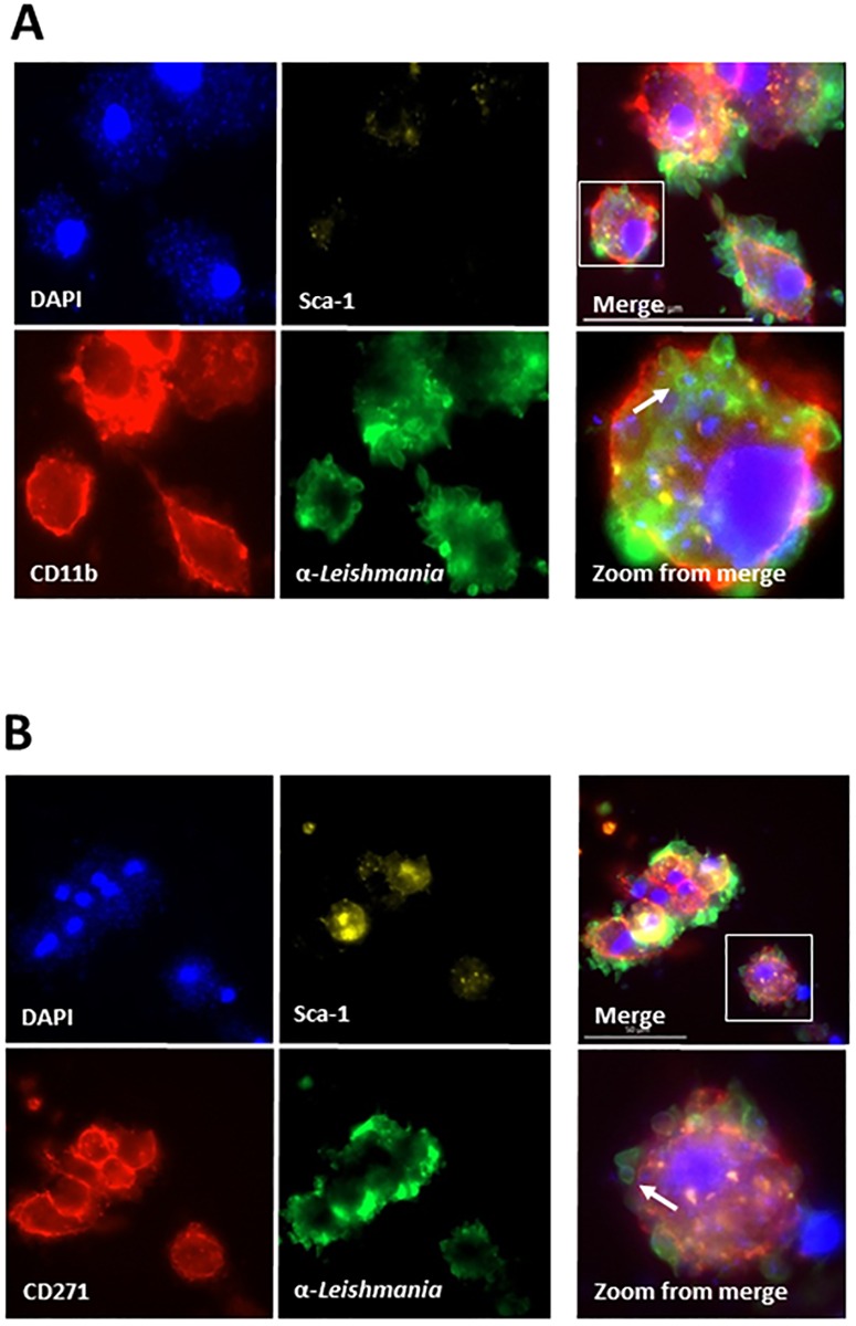
Fig 2. Confocal microscopy of mouse bone marrow mononuclear cells (BM-MNCs) co-cultured in vitro with L. infantum.
Cells were co-cultured for 96h, stained with anti-CD11b (ALEXA 568), anti-CD271+ (ALEXA 568), Sca1 (APC) and anti-Leishmania (FITC). (A), BM-MNCs cells infected with L. infantum stained with anti-CD11b, Sca1, anti-leishmania and DAPI. (B), BM-MNCs cells infected with L. infantum stained with anti-CD271, Sca1, anti-leishmania and DAPI. No staining with anti-leishmania-FITC was seen in all preparations using BM-MNCs cells not exposed to the parasites but stained with anti-CD11b, Sca1 and antiCD271 (not shown). Arrows in Zoom from merge show one of several round images that clearly suggest amastigote forms of the parasite.

