FIG 5.
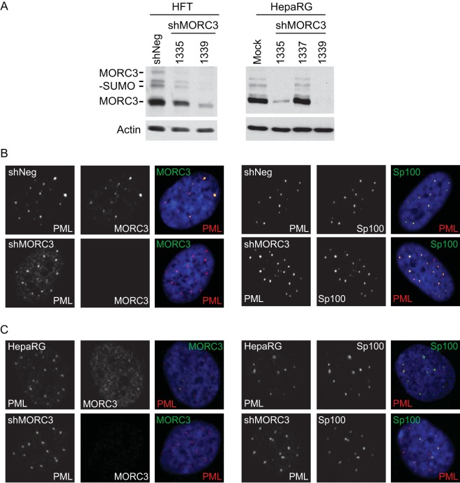
Characterization of MORC3-depleted cells. (A) Generation of MORC3-depleted HFT (HFT-shMORC3-1335 and -1339 cells) and HepaRG cells (HA-shMORC3-1335, -1337, and -1339 cells) using shRNAs expressed from lentiviral vectors. Total protein lysates were analyzed by Western blotting to determine the level of MORC3 depletion with HFT-shNeg and HepaRG cells included as controls. Tubulin was included as a loading control. (B and C) Characterization of MORC3-depleted HFT cells (B) and HepaRG cells (C). Mock HFT- and HA-shMORC3-1339 and control cells were immunostained for PML (red) and MORC3 (green) (left) as well as Sp100 (green) and PML (red) (right) to confirm MORC3 depletion and assess effects on PML NBs. DAPI staining of the nuclei is shown in blue in the merged panels.

