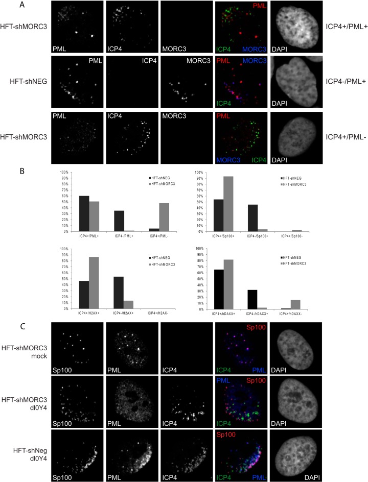
FIG 6.
Recruitment of PML NB proteins to viral DNA is diminished in the absence of MORC3. (A) HFT-shMORC3-1339 and HFT-shNeg cells were infected with dl0Y4 (MOI of 2) for 24 h. Cells were immunostained using PML (5E10) (red) and MORC3 (Novus Biologicals) (blue), and nuclei were stained with DAPI (far right) and visualized by confocal microscopy. Infected or presumed infected cells surrounding a plaque were counted and divided into three categories, which are represented here: category 1, ICP4+/PML+ (foci of ICP4 and PML in close association at the nuclear periphery of a cell); category 2, ICP4−/PML+ (foci of PML at the nuclear periphery of a cell in the pattern typical of an infected cell, but prior to detectable expression of ICP4); and category 3, ICP4+/PML− (ICP4 foci only at the nuclear periphery of a cell). (B) Percentages of cells within each category are presented in bar graphs. Sp100, hDaxx, and γH2AX were also assessed as described for PML, with results represented as bar graphs. (C) HFT-shMORC3-1339 and HFT-shNeg cells were infected with dl0Y4 (MOI of 2) for 24 h. Cells were immunostained using Sp100 (Sp26) (red) and PML (5E10) (blue) and nuclei were stained with DAPI (far right), while ICP4 was detected by the EYFP signal.

