
FIG 10.
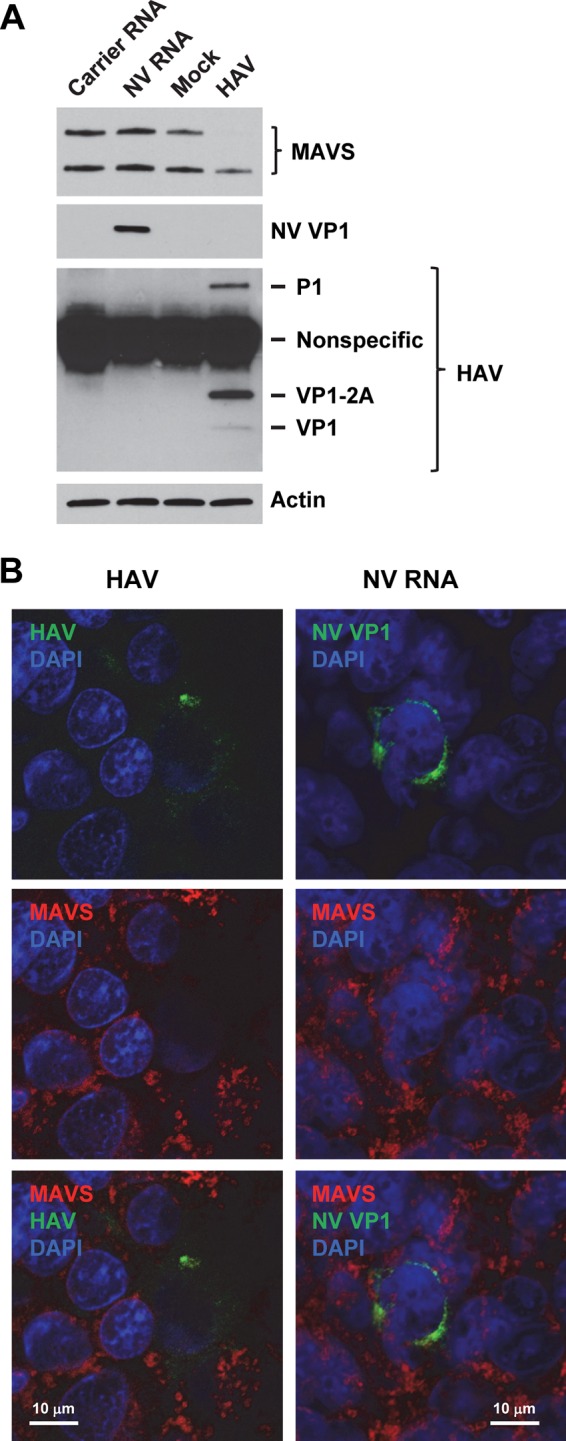
MAVS is not degraded in NV RNA replicating cells. (A) Western blotting of MAVS in 293FT cells transfected with carrier RNA or NV RNA or mock or HAV infected (multiplicity of infection of 1) for 48 h. NV RNA replication and HAV infection were confirmed by Western blotting of NV VP1 and HAV VP1/precursors, respectively. Actin served as an equal loading control. (B) Laser scanning confocal microscopy images of 293FT cells infected with HAV (multiplicity of infection of 0.1) or transfected with NV RNA for 48 h. Cells were labeled with antibodies to MAVS (red) and HAV VP1 or NV VP1 (green). Nuclei were counterstained with DAPI (blue). Scale bars, 10 μm.