
FIG 11.
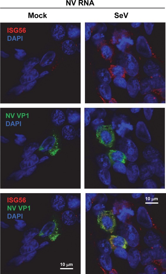
NV RNA replication does not block an SeV-induced IFN response. 293FT cells were transfected with NV RNA. At 30 h post-RNA transfection (hpt), cells were mock or SeV infected (4 HA/ml). At 48 hpt (18 hpi for SeV), cells were fixed and labeled with antibodies to ISG56 (red) and NV VP1 (green). Nuclei were counterstained with DAPI (blue). The laser scanning confocal microscopy images show that SeV-induced upregulation of ISG56 occurs in both NV VP1-positive and -negative cells. Scale bars, 10 μm.