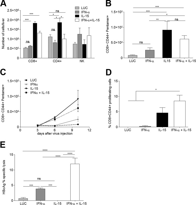
FIG 4.
IL-15-induced expansion of HBcAg-specific CD8+ T cells requires combination with IFN-α to render them functional CTLs. The mononuclear liver infiltrate was isolated and analyzed on day 10 after vector administration. (A and B) The absolute numbers of CD8+, CD4+, and NK cells per liver (A) and the number of HBcAg-specific CD8+ T cells per liver (B) are shown (n = 7). (C) The increase in the number of HBcAg-specific CD8+ T cells was analyzed over time (n = 3/time point). (D) Proliferative response of HBcAg-specific CD8+ T cells upon peptide stimulation. In vitro stimulation was performed in triplicate with cells obtained from 3 mice per group. (E) Proportion of lysed HBcAg-loaded target cells in vivo (n = 5). Results are representative of 3 independent experiments. All graphs show means ± SEM. *, P < 0.05; **, P < 0.01; ***, P < 0.001; ****, P < 0.001; ns, nonsignificant.

