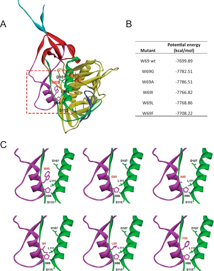
FIG 1.
Structure of HIV-1 gp120 inner domain layers 1 and 2 in the CD4-bound conformation. (A) The structure of HIV-1HXBc2 gp120 (ribbon) complexed with two-domain CD4 (omitted for clarity purposes) is shown from the approximate perspective of the Env gp trimer axis. The outer domain of gp120 is colored yellow. The N and C termini are colored cyan. The components of the gp120 inner domain are the β-sandwich (red) and three loop-like extensions: layer 1 (magenta), layer 2 (green), and layer 3 (orange). The b20-b21 strands of gp120 (blue) project from the outer domain and, in the CD4-bound conformation, are composed of two of the strands of the four-stranded bridging sheet. The other two strands of the bridging sheet are derived from the distal portion of layer 2. (B) The interaction energies of W69 variants were simulated and are listed using CHARMm implicit solvation models. (C) A closeup of the interface between layer 1 and layer 2 (red square presented in panel A) for each modeled W69 variant. The most relevant residues for layer 1-layer 2 interaction (W69, H66, D107, L111, and S115) are shown and labeled.

