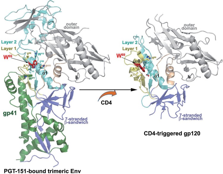
FIG 8.
Localization of W69 within the PGT151-bound HIV-1 envelope trimer and CD4-triggered gp120. The gp120-gp41 protomer from the cryo-EM structure of a fully glycosylated, PGT151-bound, cleaved HIV-1 envelope trimer (48) and gp120 coree from the Fab N5-i5-gp120 coree-d1d2CD4 structure (19) is shown as a ribbon diagram with W69 highlighted in red. Residues that contact W69 (as determined by 4-Å distance cutoff) are shown as a ball and sticks.

