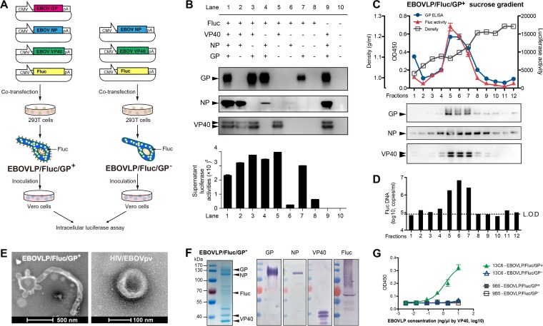
FIG 1.
Construction and characterization of the reporter EBOVLP. (A) Schematic diagram of the generation of complete EBOVLP/Fluc/GP+ and envelope-lacking EBOVLP/Fluc/GP−. (B) Expression of GP, NP, VP40, and Fluc in the supernatant of HEK293T cells transfected with plasmids in different combinations. EBOV proteins GP, NP, and VP40 were identified by Western blotting, while the presence of Fluc was detected by luciferase assay. (C and D) Sucrose gradient analysis of cell culture supernatant containing EBOVLP/Fluc/GP+. The presence of EBOV proteins in the collected fractions was detected by ELISA and Western blotting (C), while the distribution of Fluc protein and Fluc plasmid was tested by luciferase assay (C) and qPCR (D). The dashed line indicates the limit of detection (L.O.D). (E) Electron microscopy of purified EBOVLP/Fluc/GP+ and HIV/EBOVpv. (F) SDS-PAGE and Western blot analyses of purified EBOVLP/Fluc/GP+. The band(s) of each protein was verified by Western blotting using corresponding primary antibodies and alkaline phosphatase-conjugated secondary antibodies. (G) Recognition of EBOVLP/Fluc/GP+ by an EBOV-neutralizing MAb, 13C6, but not a control MAb, 9B5, in ELISA. Means ± standard errors of the mean (SEM) of the optical density at 45 nm (OD450) readings are shown.

