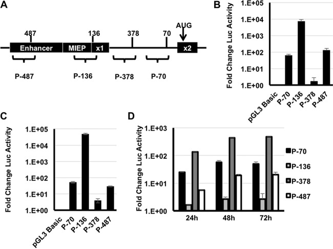
FIG 4.
DNA sequences surrounding the novel MIE transcription start sites have promoter activity. (A) Cartoon showing the location of the 500 nucleotide regions tested for promoter activity. (B) HeLa cells were transfected with the indicated reporter plasmids, and luciferase activity was measured at 24 h after transfection. The graph shows the fold change in luciferase (Luc) activity relative to cells transfected with the empty pGL3 Basic vector, which lacks a promoter for the luciferase gene. (C) The indicated reporters were electroporated into MRC-5 fibroblasts, and luciferase activity was measured at 24 h after electroporation. (D) MRC-5 fibroblasts were electroporated with the indicated reporters and then infected with HCMV (MOI of 3). Luciferase activity was measured at the indicated times after HCMV infection. The graph shows the fold change in luciferase activity relative to the activity of each reporter at 6 h after infection. P-, promoter.

