FIG 5.

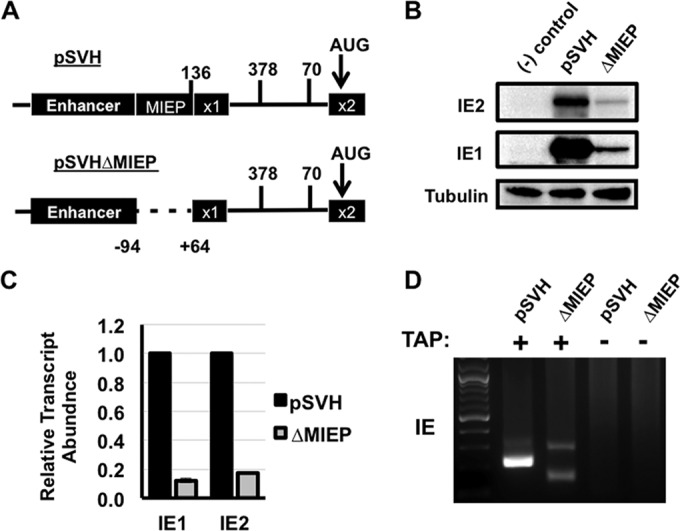
The core promoter region of the MIEP is not necessary for IE1 and IE2 expression outside the context of HCMV infection. (A) Cartoon showing a portion of the MIE locus in pSVH. The numbers show the locations of the MIE transcription start sites for the indicated MIE transcripts shown in Fig. 1. pSVHΔMIEP was created by removing a 158-bp region containing the MIEP core promoter (−94 to +64 relative to the transcription start site). (B) HeLa cells were left untransfected {negative control [(-) control]} or transfected with pSVH or pSVHΔMIEP (ΔMIEP). IE1 and IE2 protein levels were measured by Western blotting at 24 h after transfection. (C) HeLa cells were transfected and harvested as in panel B. The relative abundance of the IE1 and IE2 mRNAs in pSVHΔMIEP-transfected cells compared to pSVH-transfected cells was determined by qRT-PCR using the ΔΔCT method. IE1 and IE2 abundance in pSVH-transfected cells is set at one. (D) HeLa cells were transfected as in panel B. 5′ RACE analysis of polysome-associated RNA was performed using gene-specific primers located in exon 2. 5′ RACE PCR products were visualized on agarose gels and subsequently cloned and sequenced. No PCR products were obtained in cells where tobacco acid pyrophosphatase (TAP) was omitted (−), demonstrating that the PCR products were derived from mRNAs containing a 5′ m7G cap.
