FIG 6.
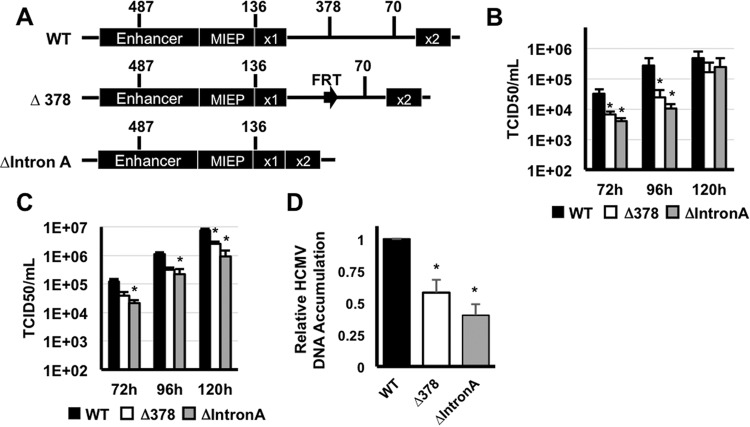
Removal of MIE intron A delays HCMV replication. (A) Diagram depicting the genomic regions removed from each recombinant HCMV BAC. (B) MRC-5 fibroblasts were infected with wild-type (WT) HCMV, HCMVΔIntron A (ΔIntron A), or HCMVΔUTR378 (ΔUTR378) (MOI of 0.5). Cell-free virus was quantified at the indicated times after infection by the TCID50 assay. (C) Fibroblasts were infected with HCMV (MOI of 3), and cell-free virus was quantified as in panel B. Values that are significantly different (P < 0.05) from the WT value are indicated by an asterisk. (D) Fibroblasts were infected as in panel B, and the relative abundance of viral DNA was quantified by real-time PCR at 72 h after infection. The amount of viral DNA in cells infected with wild-type virus was set at one. Values that are significantly different (P < 0.001) from the WT value are indicated by an asterisk.

