FIG 1.
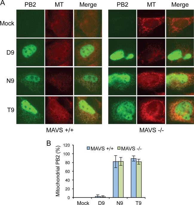
MAVS is not required for the mitochondrial localization of PB2. (A) Cellular distribution of GFP-tagged PB2 with D9, N9, or T9 in transfected MAVS+/+ and MAVS−/− MEF cells. Cells were stained with the fluorescent mitochondrial marker Mitotracker (MT) and imaged by fluorescence microscopy. (B) PB2-expressing cells were scored for mitochondrial localization of PB2. Column data represent the percentages of PB2-expressing cells with mitochondrial PB2 signal. Bars represent standard deviations of averages of data from three independent experiments (n = 17 to 21 cells/experiment).

