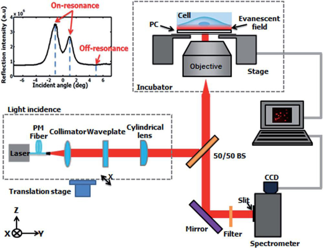
Fig. 14.
Overview of the detection instrumentation, composed of a laser light source directed into a microscope objective enclosed in an incubation chamber. The incident angle is tuned via the translation stage. The sample stage holding the PC also translates along the axis perpendicular to the imaged line for each scan. Scans are performed in 0.6 µm increments, at a rate of 0.1s per line to form a whole image. Reprinted with permission from [47], © 2014 RSC Publishing.

