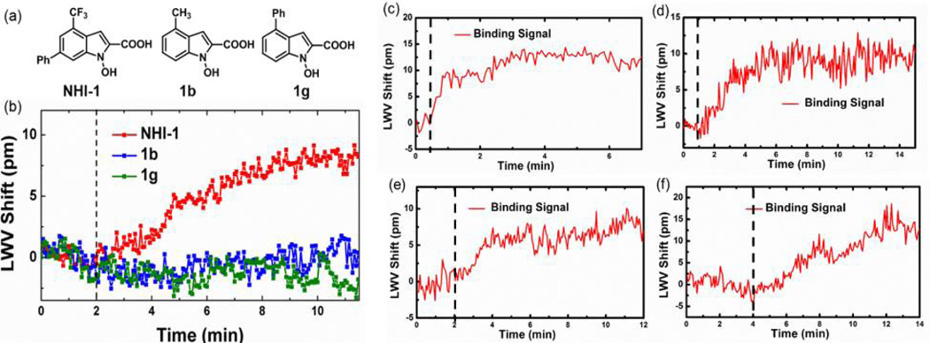
Fig. 2.
(a) Structure of NIH-1 and two inactive variants 1b and 1g. Observed LWV shift from the binding of 50 µM (b) NHI-1, 1b and 1g to immobilized hLDH-A, (c) SM-164 to immobilized GST-XIAP, (d) dorzolamide to immobilized CA-II, (e) dicoumarol to immobilized NQO1, and (f) Q-VD-OPh to immobilized caspase-3. Dashed line represents the time at which small molecule was added to the active well. Self-referencing was applied to each. Reprinted with permission from [82], © 2014 American Chemical Society.

