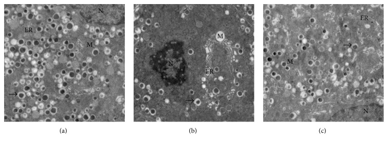
Figure 11.
Electron microscopic analysis of pancreatic beta cells in different groups. (a) Ultrastructure of pancreatic beta cell showed that the nuclei, endoplasmic reticulums, mitochondria, and secretory granules were not abnormal in SO group (original magnification, ×3,000). (b) The changes of pancreatic beta cells in SAP group show condensed chromatin and loss of secretory granules, concomitant with severely dilated endoplasmic reticulums and swollen mitochondria (original magnification, ×3,000). (c) Mild dilatation of the endoplasmic reticulums, decrease of mitochondria damage, and increase of secretory granules were noted in 4-PBA group (original magnification, ×3,000). Note. Nucleus (N), mitochondria (M), endoplasmic reticulum (ER), and secretory granules (black arrow).

