. 2016 Sep 13;5:e18605. doi: 10.7554/eLife.18605

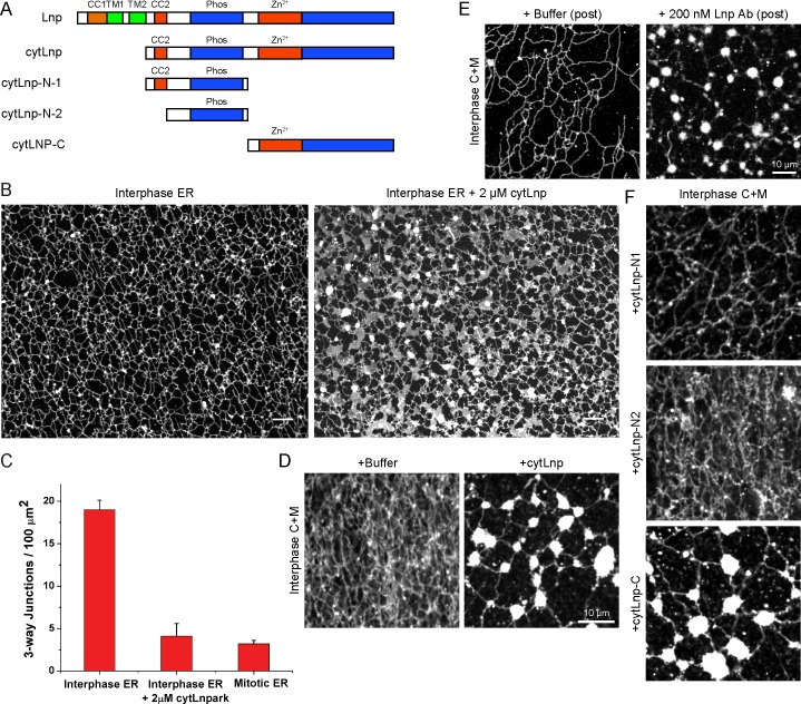
Figure 11. Effect of Lnp inactivation on an in vitro generated ER network.

(A) Schematic representation of wild type and mutant Xenopus Lnp. Phos indicates the domain phosphorylated during mitosis. (B) An ER network was generated for 20 min with crude Xenopus egg extract in the presence of the dye DiIC18 and either buffer (left panel) or 2 µM of a cytoplasmic fragment of Lnp (cytLnp). Scale bar = 10 µm. (C) The number of three-way junctions at different conditions was quantitated. Error bars indicate the mean ± SD of three independent experiments. (D) An interphase ER network was formed with Xenopus egg cytosol (C), light membranes (M), an energy regenerating system, in the presence or absence of 5 µM cytLnp. The network was stained with octadecyl rhodamine. Scale bar = 10 µm. (E) Buffer or 200 nM of affinity-purified Lnp antibodies were added to a preformed interphase network generated with cytosol, membranes, and an energy regenerating system. Scale bar = 10 µm. (F) As in (D), except that 5 µM cytLnp-N1, cytLnp-N2 or cytLnp-C were added at the beginning of the network formation reaction. Scale bars = 10 µm.

DOI: http://dx.doi.org/10.7554/eLife.18605.027

Figure 11.

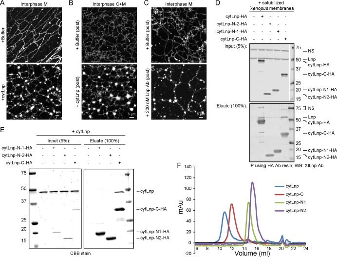
Figure 11—figure supplement 1. Cytoplasmic fragments of Lnp interfere with the function of endogenous Xenopus Lnp.

Figure 11—figure supplement 1.

(A) Light membranes (M) were incubated with an energy regenerating mix and either buffer or 5 µM cytLnp for 15 min. The membranes were stained with octadecyl rhodamine. Scale bar = 5 µm. (B) Buffer or 5 µM of cytLnp was added to a preformed network formed from cytosol (C) and membranes (M). Scale bar = 5 µm. (C) Buffer or 200 nM of affinity-purified Lnp antibody was added to a preformed network formed from membranes only. Scale bar = 5 µm. (D) Interaction of endogenous Xenopus egg Lnp with various Lnp fragments. A detergent extract of light membranes was incubated with empty resin or hemagglutinin (HA)-tagged Lnp fragments (see Figure 11A). After pull-down with HA antibodies, the bound fractions were analyzed by immunoblotting with antibodies to Xenopus Lnp. (E) Interaction of purified Xenopus cytLnp with various cytLnp fragments. HA-tagged Lnp fragments (see Figure 11A) was incubated with cytLnp, and subsequently HA antibodies. Bound fractions were analyzed by SDS-PAGE and stained with Coomassie blue. (F) Purified cytLnp or fragments of it (see Figure 11A) were analyzed by gel filtration on a Superdex 200 column.