. 2016 Sep 13;5:e18605. doi: 10.7554/eLife.18605

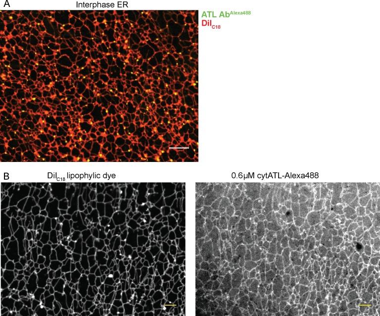
Figure 5. Localization of ATL in an in vitro generated ER network.

(A) An interphase ER network was generated with a crude Xenopus egg extract containing the dye DiIC18. Endogenous ATL was visualized by including 16 nM Alexa488-labeled, affinity-purified antibodies raised against Xenopus ATL (ATL AbAlexa488). Note that ATL localizes preferentially to three-way junctions. Scale bar = 10 µm. (B) An interphase ER network was assembled as in (A) and labeled with DiIC18 and 0.6 µM Alexa488-labeled cytATL. Note that cytATL localizes throughout the tubules, marking the position of inactivated endogenous ATL. Scale bar = 10 µm.

DOI: http://dx.doi.org/10.7554/eLife.18605.014

Figure 5.

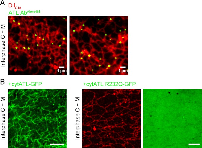
Figure 5—figure supplement 1. Localization of endogenous and inactivated ATL in a Xenopus ER network.

Figure 5—figure supplement 1.

(A) An interphase ER network was generated with cytosol, DiIC18-prelabeled light membranes, and an energy regenerating system. Endogenous ATL was visualized by including 10.5 nM Alexa488-labeled, affinity-purified antibodies against Xenopus ATL. The sample also contained sperm chromatin to stabilize the network by reducing thermal convection. Scale bars = 1 µm. (B) A mixture of interphase cytosol, light membranes, and an energy regenerating system was incubated in the presence of 0.5 µM cytATL-GFP, but absence of membrane stain,for 30 min (left panel). Parallel reactions were performed with 2 µM cytATL(R232Q)-GFP and the membrane stain octadecyl rhodamine (middle and right panels). Scale bars = 10 µm.