. 2016 Sep 13;5:e18605. doi: 10.7554/eLife.18605

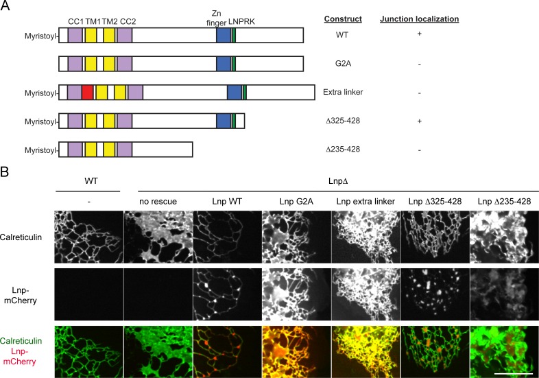
Figure 9. Lnp domains important for junction localization.

(A) Schematic representation of wild type (WT) Lnp and mutants tested for proper localization in the peripheral ER. CC1, CC2, coiled-coil domains 1 and 2, respectively; TM1, TM2, trans-membrane segments 1 and 2, respectively; LNPRK, lunapark motif (single letter code); the segment indicated in red was inserted (extra linker). (B) Peripheral ER in U2OS cells expressing GFP-calreticulin under the endogenous promoter in wild type or Lnp-lacking cells (LnpΔ). Where indicated, the cells also stably expressed wild type or mutant Lnp at low levels. Scale bar = 10 µm.

DOI: http://dx.doi.org/10.7554/eLife.18605.023

Figure 9.

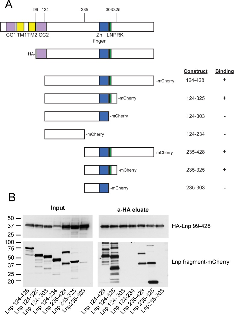
Figure 9—figure supplement 1. Lnp-Lnp interaction is mediated by the Zn2+-finger domain-containing region.

Figure 9—figure supplement 1.

(A) Schematic representation of Lnp truncation mutants tested for interaction. Hemagglutinin (HA)-tagged Lnp 99–428 was used as bait for pull-downs of mCherry-tagged truncations. (B) Immunoblots of co-immunoprecipitation experiments. Each of the mCherry-tagged fragments and the HA-tagged bait were expressed separately in 293T cells. Cells were lysed, and the levels of mCherry-tagged truncations were normalized according to fluorescence at 590 nm, except Lnp 235–303 which was poorly expressed. After mixing with lysate containing HA-Lnp 99–428, pull-downs were performed with HA-antibodies and the samples were analyzed by immunoblotting with HA- and mCherry- antibodies.