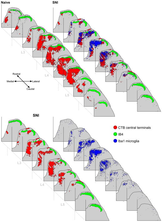
Fig. 5.
Top panel: Reconstructions of naïve and SNI lumbar dorsal horns showing the somatotopic nature of the microglial response to PNI. Microglia are indicated in blue, CTB in red and IB4 in green. Bottom panel: SNI dorsal horn with the microglial labelling (right) separated from the primary afferent labelling (left). While the majority of microglial activity is restricted to the injured central terminal fields, shown by overlap of the blue and red labelling, there is a small but distinct spread of microglial labelling into the intact sural territory. (For interpretation of the references to color in this figure legend, the reader is referred to the web version of this paper.)

