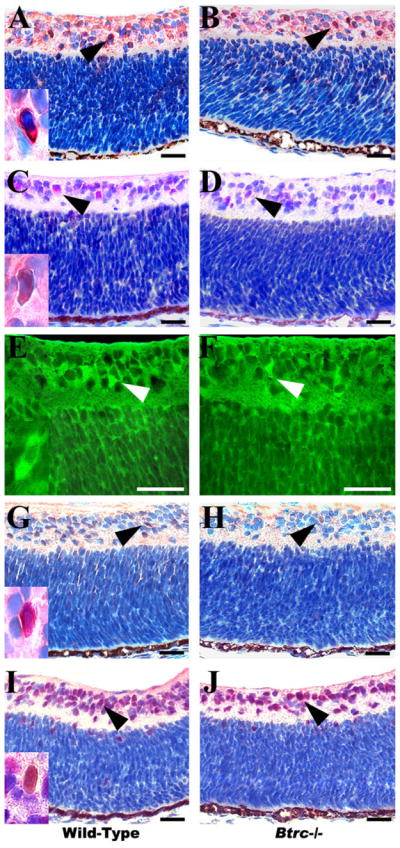
Fig. 4.

Aberrant amacrine and ganglion cell numbers in the Btrc−/− retina. Immunohistochemistry against the cytoplasmic markers calretinin (A,B), calbindin (C,D), parvalbumin (E,F), and tyrosine hydroxylase (G,H), (arrowheads) reveals a decrease in tyrosine hydroxylase-expressing amacrine cells in the Btrc−/− retina (H), whereas the population of other amacrine cell subtypes appears similar to that in the wild-type retina. The number of retinal ganglion cells, indicated by the nuclear marker Islet 1/2, was also lower in the mutants (J) than in the wild-type (I). Insets are higher magnifications of positively stained cells. Scale bar = 25 μm.
