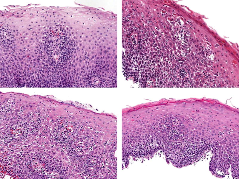
Figure 2.

Histologic findings of patients with lymphocytic esophagitis showing esophageal squamous mucosa with variable spongiosis and increased numbers of intraepithelial lymphocytes in a diffuse distribution, ranging in numbers from mild to striking, occasionally forming small lymphocytic clusters, particularly in the peripapillary areas.
