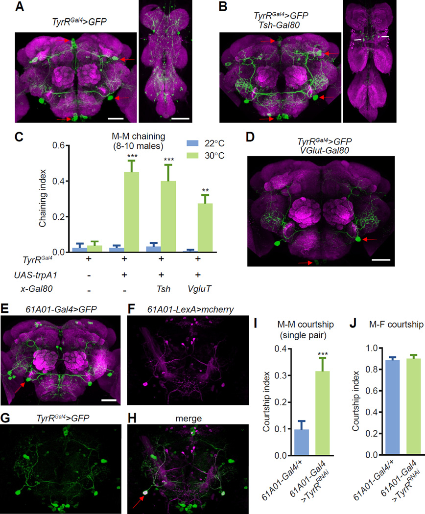
Figure 4. TyrR controls courtship behavior through IPS neurons.
(A) Staining of an adult male brain (left) and VNC (right) using the TyrR-Gal4 reporter (TyrRGal4/+) in combination with UAS-mCD8-GFP. Anti-GFP (green) and anti-nc82 (panneuropil marker, magenta).
(B) Elimination of TyrR-reporter expression in most cells in the VNC using a Gal4 repressor (Gal80): Tsh::Gal80. Anti-GFP (green) and anti-nc82 (pan-neuropil marker, magenta).
(C) Male chaining resulting from thermal hyperactivation of defined subsets of TyrR-expressing neurons using two Gal80 transgenes. To determine statistical significance, we compared the chaining indices at 22°C (no TRPA1 activation) and 30°C (TRPA1 activated). n=4–7 trials/genotype.
(D) VGlut::Gal80 restricted TyrRGal4 expression to the IPS and GNG regions of the male brain. The image was obtained from an animal maintained at room temperature.
(E) UAS-mCD8-GFP expressed under the control of the 61A01-Gal4. The red arrow indicates the IPS neurons in the brain. Anti-GFP, green; anti-nc82, magenta.
(F-H) IPS neurons co-expressed the TyrRGal4 and the 61A01-LexA. The brains contained the following transgenes: 61A01-LexA/UAS-mCD8-GFP;TyrRGal4/LexAop2-mCherry. Anti-GFP, green; anti-DeRed, magenta.
(I) Male-male courtship indices after RNAi knockdown of TyrR in 61A01-Gal4 neurons. n=20–22.
(J) Male-female courtship indices after RNAi knockdown of TyrR in 61A01-Gal4 neurons. n=15–19.
The immunohistochemistry experiments were performed at room temperature. The scale bars represent 50 µm. The error bars indicate the means ±SEMs. Mann-Whitney test were performed. **p<0.01. ***p<0.001.

