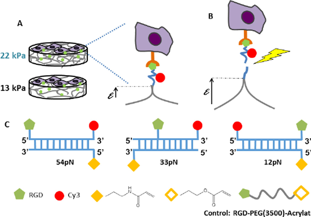
Figure 1.
Schematic of the force response of TGTs on soft substrates. (A) Cell attachment to TGT-modified hydrogels with elastic moduli of 13kPa (soft) and 22kPa (rigid). (B) Cells deform the substrates until the tension exceeds tension tolerance (Ttol) and the cells tear off the sensing strand (yellow jagged arrow). (C) DNA duplex tethers with tunable Ttol. The anchor DNA is covalently linked to the gel through polymerizable acrydite monomers (solid yellow diamond). The ‘reporter’ strand contains a 3’ Cy3 dye (red circle) and the location of the RGD peptide along the strand (green pentagon) determines the tension tolerance Ttol. In this study, Ttol values were 12pN, 33pN, or 54pN. The control is a RGD peptide covalently linked to an acrylate group (hollow yellow diamond).

