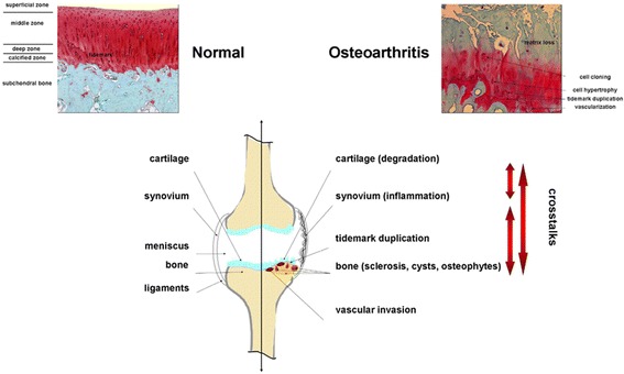
Fig. 1.

OA-associated changes in the human joint. Safranin O-stained histological sections from human normal versus OA knee joint cartilage are presented on the top panels. The lower panel is a representation of the different tissues implicated in the OA pathology with crosstalks
