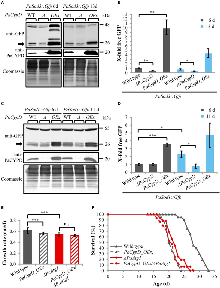
Figure 2.
Autophagy-dependent degradation of mitochondrial and cytosolic marker proteins in wild-type strain and different PaCypD mutants during aging. (A) Monitoring mitophagy by western blot analysis using the mitochondrial protein PaSOD3::GFP. 6 and 13 d old wild-type (WT), ΔPaCypD and PaCypD_OEx strains expressing PaSod3::Gfp were cultured for 2 d in CM medium. “Free GFP” (indicated by arrow) and PaCYPD were monitored by immunoblotting with anti-GFP and anti-PaCYPD in 100 μg total protein extracts. The positions of molecular mass markers are indicated on the right. (B) The GFP protein levels of three different isolates of each strain were quantified and normalized to the Coomassie-stained gels (loading control). The protein amount present in the 6 d old wild type was set to one. (C) Monitoring autophagy as described in (A) except using 6 and 11 d old strains expressing PaSod1::Gfp. (D) Quantification of “free GFP” derived from the cytosolic marker PaSOD1::GFP as described in (B). (E) Growth rate and (F) lifespan of wild type (n = 20), PaCypD_OEx (n = 20), ΔPaAtg1 (n = 16), and the double mutant PaCypD_OEx/ΔPaAtg1 (n = 15) on M2 medium. Data represent average ± SEM (2-tailed student's t-test), *P < 0.05, **P < 0.01, ***P < 0.001.

