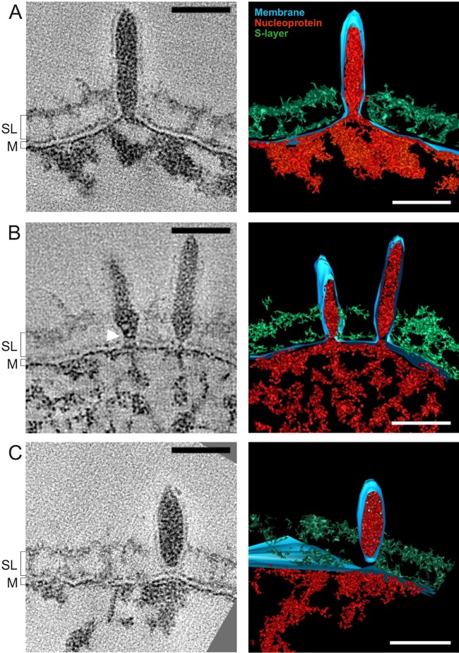
FIG 1 .
Different stages of SSV1 budding. (A to C) Slices through tomograms (left) and volume segmentations (right) showing concomitant assembly and release of SSV1 virions (see Videos S1 and S2 in the supplemental material). The white arrowhead marks an electron density presumed to be a ring-like structure. Red, putative nucleoprotein; blue, lipid membrane (M); green, S-layer (SL). Scale bars, 50 nm.

