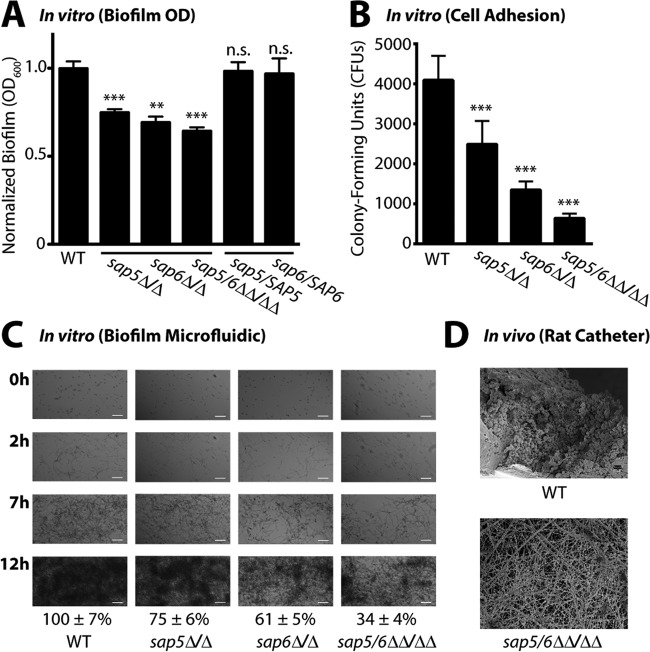
FIG 6 .
Deletion of SAP5 and SAP6 impairs C. albicans biofilm formation under in vitro growth conditions and in vivo in a rat central venous catheter biofilm model. (A) Biofilm formation in Spider medium after 24 h of growth of the wild-type (WT) reference (SN250) and sap5Δ/Δ, sap6Δ/Δ, and sap5/6ΔΔ/ΔΔ deletion strains. OD600 readings were measured for adhered biofilms after removal of the medium and normalized to the wild-type strain (OD600 set to 1.0), and the mean ± SD is shown (n = 4 for each strain). OD600 measurements of the sap5, sap6, and sap5/6 deletion strains deviated significantly from those of the reference strain (**, P < 0.01; ***, P < 0.001; n.s., not significant). Complementation of the sap5Δ/Δ and sap6Δ/Δ deletion mutant strains with SAP5 and SAP6, respectively, restored biofilm formation to wild-type reference levels, with P = 0.70 for sap5/SAP5 and P = 0.66 for sap6/SAP6 (P values were calculated by comparison to the reference strain). The growth rates of the sap5 and sap6 deletion strains in the planktonic state were not compromised, as shown in Fig. S8 in the supplemental material. (B) Quantification of early adhered cells under the biofilm assay condition in Fig. 6A immediately following the 90-min adherence period (n = 12 for each strain). The CFU counts (reported as mean ± SD) of the sap5Δ/Δ, sap6Δ/Δ, and sap5/6ΔΔ/ΔΔ deletion strains deviated significantly from those of the reference strain (***, P < 0.05 by one-way ANOVA). (C) Time-dependent visualization of biofilm formation under dynamic flow (0.5 dyne/cm2) in Spider medium over a 12-h period postadherence with a BioFlux 1000z instrument, with representative 0-, 2-, 7-, and 12-h images shown (n = 3 for each strain). Bars = 50 µm in all panels. Areas occupied by the mature biofilms at 12 h were normalized to the wild-type (SN250) reference strain. Normalized percent areas (reported as mean ± SD) of the sap5Δ/Δ(75 ± 6%), sap6Δ/Δ(61 ± 5%), and sap5/6ΔΔ/ΔΔ (34 ± 4%) deletion strains deviated significantly (P < 0.01) from those of the reference strain (100 ± 7%). Corresponding time-lapse videos of biofilm formation are provided in Movie S1 in the supplemental material. (D) Comparison of in vivo biofilm formation by the C. albicans wild-type reference (SN250) and sap5/6ΔΔ/ΔΔ deletion strains in the rat central venous catheter biofilm model. SEM images acquired at 24 h postadherence illustrate a reduction in the number of adhered cells of the C. albicans sap5/6ΔΔ/ΔΔ deletion strain (host protein deposited on the intraluminal surface is evident). Representative images of biological duplicates are shown at ×1,000 magnification. White bars, 10 µm. The sap5/6ΔΔ/ΔΔ deletion did not affect planktonic virulence in vivo as shown in Fig. S8 in the supplemental material.

