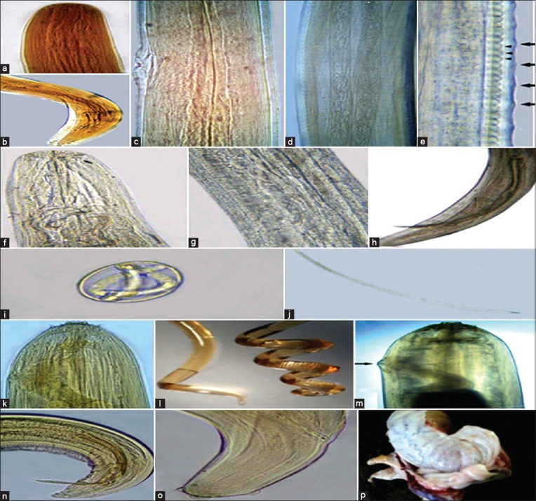
Figure-1.

Onchocerca cervicalis: (a) Anterior end, (b) male posterior end, (c) vulva, (d) microfilariae in the uterus of female (e) middle region of female showing low widely spaced indistinct external cuticular annulations (large arrows) and internal striations forming elongated cells (small arrowheads); Parafilaria multipapillosa: (f) Anterior end (arrow refers to vulva anteriorly near the mouth opening), (g) Uteri in the middle region, (h) male posterior end, (i) embryonated egg with thin flexible shell, (j) microfilaria with rounded posterior extremity; Setaria equina: (k) Anterior end, (l) coiled male posterior end (right) and female (left), (m) female vulva, (n) male posterior end, (o) female posterior end, (p) adult worm encapsulated between the testicular tunica.
