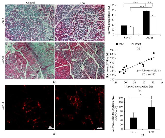
Figure 6.
Histologic specimens obtained from ischemia muscle. (a) At days 3 and 28, almost complete tissue recovery using Masson's staining was found in the EPCs treated group. However, in the control group, multiple inflammations and collagen were found (bar = 100 μm). (b) A bar graph showed that the survival muscle fibers of mice treated with EPCs were greater than with the control group. Data is expressed as mean ± SD, ∗ P < 0.05. (c) A line graph showed the correlations between survival muscle fibers by histological method and fiber count measured by DTI (r = 0.874, P < 0.01). (d) The microvascular density was measured by staining ischemia tissues with CD31 on day 14 after treatment. The EPC treatment augmented vessel density of the ischemic hindlimb (bar = 30 μm). (e) A bar graph showed that the microvascular density (MVD) in the group treated with EPCs was significantly increased compared with that of the control group. ∗ P < 0.05, ∗∗ P < 0.01, and ∗∗∗ P < 0.001.

