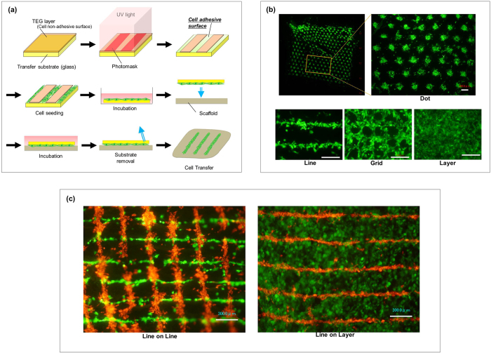
Figure 1. Cell transfer technology and double cell transfer.
(a) Schematic diagram of cell transfer technology. We coated the surface of glass substrate with tetraethyleneglycol (TEG, brown) and the layer was partially degraded by UV irradiation to prepare hydrophilic cell adhesive surface (green). The TEG area covered with photomask remained as non-cell adhesive surface. Cells to be transferred were poured onto the substrate and incubated to allow the cells to adhere to the substrate surface. Transfer substrate with cells was then placed onto the scaffold (amnion) in the direction of cell surface down. Cells were further cultured and transfer substrate was carefully removed subsequently. Cells are transferred onto scaffold surface. (b) Various cell patterning by using cell transfer technology. PDLSC (GFP; green) were transferred onto amnion using patterning base for dot, line, grid and layer placement. Boxed area was closed-up on the right figure. Bar = 300 μm (c) Double cell transfer using line patterning and layer transfer base. Fibroblasts (PKH26; red) were transferred using line patterning base and PDLSC (GFP; green) were transferred overlaying the fibroblasts displacing 90 degrees (left). PDLSC in layer structure was transferred on fibroblasts in line pattern (right). Bar = 300 μm.

