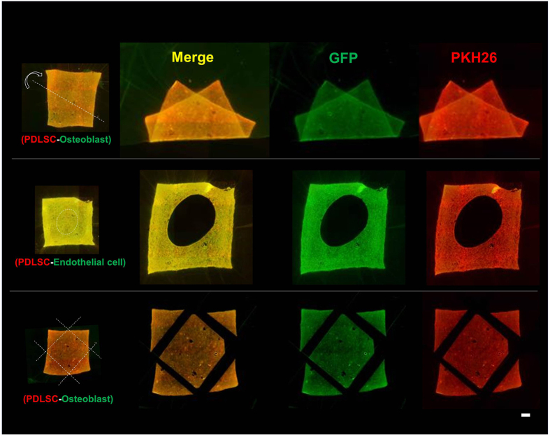
Figure 4. Stability of transferred cells upon trimming and deformation of amnion.
Fluorescence microscopic images of amnion holding double-layered cells after deformation (top), holing (middle) and trimming (bottom) of the membrane. After double-layered cell transfer, using PDLSC (GFP, green) and KUSA-A1 (PKH26, red), amnion was folded along the dotted line (top). Circular hole was made in the middle of cell-transferred amnion (middle). We used PDLSC (GFP, green) and HUVEC (PKH26, red) for double cell transfer. Four corners were trimmed along by the dotted line after double-layered cell transfer (bottom). Despite of deformations and trimming of cell transferred amnion, cells were stably adhered onto the scaffold material. We used PDLSC (GFP, green) and KUSA-A1 (PKH26, red) for double cell transfer, except for holing. PDLSC was used for the first layer in all cell transfer tested.

