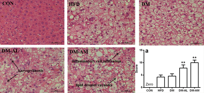
Figure 2. Histological analyses of livers from normal rats (CON), rats fed with high-fat diet (HFD), diabetic rats (DM), diabetic rats treated with 10 mg/kg atorvastatin (DM-AL) and 20 mg/kg atorvastatin (DM-AM).
All sections were stained with hematoxylin-eosin (magnification, 200×). Scale bar represented 500 μm. Histopathological damage scores for all experiment rats (a). Data were represented as means ± SD (n = 5). *p < 0.05, **p < 0.01 vs. DM.

