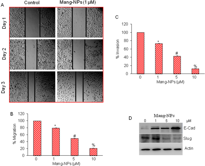
Figure 5. α-mangostin inhibits cell motility, migration and invasion and modulates the expression markers of epithelial-mesenchymal transition (EMT).
(A) Pan CSCs were grown in monolayer, scratched and treated with or without Mang-NPs (0–1 μM) for 1 or 2 days. Cells were photographed as we described elsewhere22,50. (B,C) Cell Migration and invasion assay. Pan CSCs were seeded, treated with Mang-NPs (0–10 μM) for 48 h and cell migration and invasion assays were performed as described in Materials and Methods. Data represent mean (n = 4) ± SD. *, #, and % = significantly different from control, and each other, P < 0.05. (D) Pan CSCs were treated with Mang-NPs (0–10 μM) for 48 hrs. The expression of E-cadherin, and Slug was measured by the Western blot analysis. β-actin was used as an internal control.

