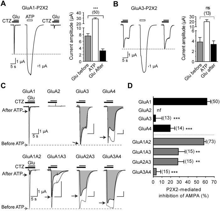
Figure 1. P2X2-mediated inhibition of AMPAR current is dependent upon AMPAR subunit composition.
(A,B) Representative currents evoked by application of glutamate (Glu 1 mM for 5 s) in the presence of cyclothiazide (CTZ, 100 μM, 10 s of preincubation) before and 2 min after an ATP-induced current (100 μM) in oocytes co-expressing P2X2 and either GluA1 (A) or GluA3 subunits. (B) Summary of amplitude averages of AMPAR and P2X2 currents. (C) Superimposed AMPAR currents evoked in the same conditions as in A,B before (gray traces, unfilled areas) and 2 min after an ATP-induced current (black traces and shaded areas) for oocytes expressing P2X2R and indicated homomeric or heteromeric AMPARs. (D) Bar graphs summarizing the extent of inhibition (expressed as %) of homomeric or heteromeric AMPAR after activation of P2X2R. Statistical differences compared to GluA1 or GluA1A2 are indicated. **P < 0.01; ***P < 0.001; ns, P > 0.05; Error bars represent s.e.m.; Numbers of cells are indicated in parentheses. nf, non-functional.

