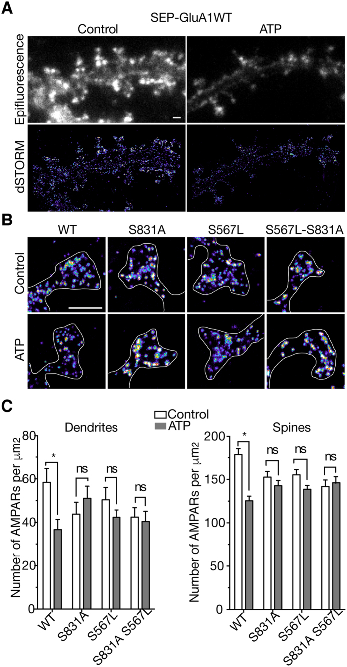
Figure 5. Decrease in number of dendritic and synaptic SEP-tagged GluA1 receptors triggered by activation of native P2XR in transfected hippocampal neurons is mediated by the S831 or S567 GluA1 residues.
(A) Epifluorescence (upper panels) and super-resolution images (bottom panels) reconstructed from direct Stochastic Optical Reconstruction Microscopy (dSTORM) of wild-type (WT) SEP-tagged GluA1 in transfected hippocampal neurons labeled with surface anti-GFP antibodies before (control, left panel) and after ATP treatments (right panel). (B) Representative dSTORM images of spines from neurons expressing wild-type SEP-tagged GluA1 and mutant GluA1 S831A, S567L and double mutant in control conditions or 1a0 min after application of ATP (100 μM, 1 min) in presence of CGS15943 (3 μM) and TTX (0.5 μM). Scale bars, 1 μm. (C) Average density values of wild-type and mutant SEP-GluA1-containing AMPAR in synapses and dendrites in control condition (unshaded bars) and after P2XR activation (shaded bars). *P < 0.05; ns, P > 0.05; Error bars: s.e.m.

