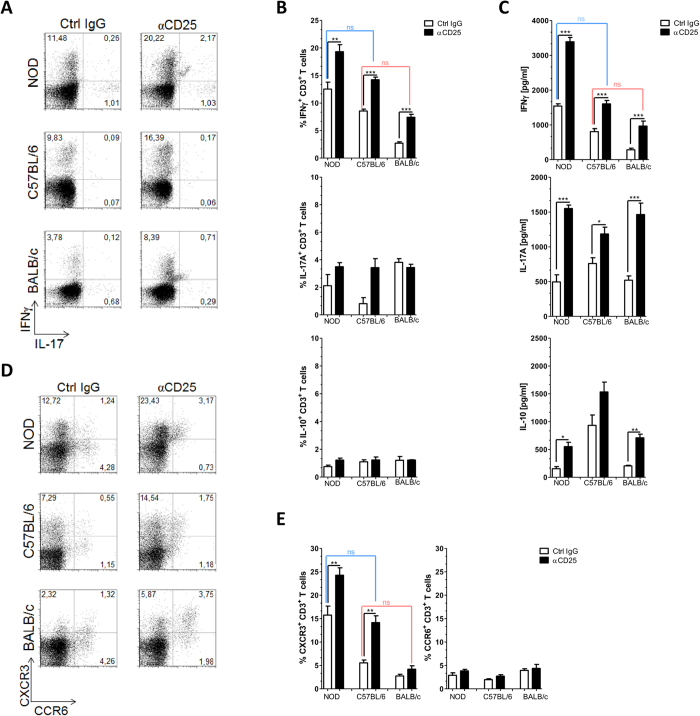
Figure 2. CD25+ cell depletion diverts specific immune response towards a Th1/Tc1 pattern.
Mice were treated as in Fig. 1, pooled draining LN from individual NOD, C57BL/6 and BALB/c mice were analyzed at day 24, either straight (A) or after stimulation with PSBP for 72 h (B–E). (A) Flow cytometric analysis for IFNγ and IL-17A intracellular expression in CD3+ cells isolated from draining LN following in vitro stimulation with PMA/inomycin for 5 h in the presence of Golgi Stop. (B) Percentage of IFNγ+ CD3+, IL-17A+ CD3+ and IL-10+ CD3+ T lymphocytes isolated from draining LN following in vitro stimulation with PSBP for 72 h and then 5 h. PMA/ionomycin incubation in the presence of Golgi Stop as described in Materials and Methods. (C) Culture supernatants of mononuclear cells from draining LN isolated on day 24 of the experimental protocol, analyzed by ELISA. (D) Representative flow cytometry dot plots of gating strategy and CXCR3 and CCR6 staining on the gated CD3+ T cell population after 72 h of PSBP stimulation. (E) Percentages of CXCR3+ and CCR6+ in CD3+ T cells after 72 h of PSBP stimulation. Data are shown as mean ± SEM, n = 6 per group, and are representative of two independent experiments. The p values were obtained using one-way ANOVA followed by Bonferroni post-hoc analysis; ns, non significant.

