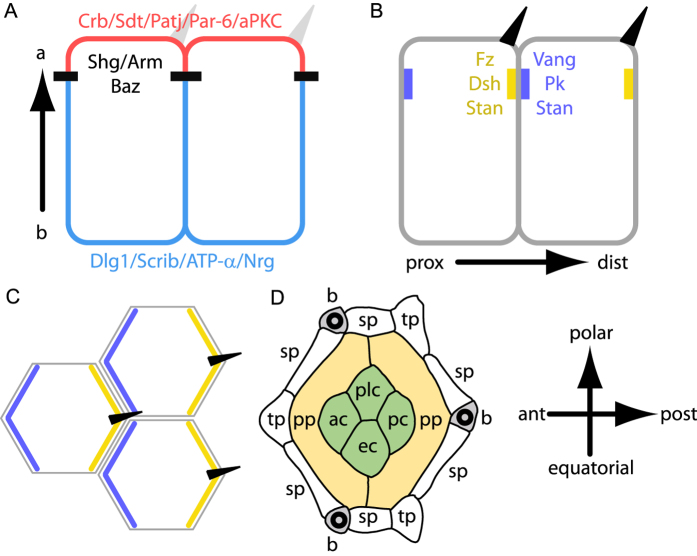
Figure 1. Planar and transversal distributions of apico-basal and planar polarity proteins.
(A,B) transverse section showing the distribution of apico-basal (A) and planar polarity proteins (B) in fly wing epithelial cells. (A) The apical most region of the cell is shown in red, adherens junction are indicated in black and baso-lateral domains in blue. (B) Proximal (prox) PCP domains containing Vang, Pk and Stan are indicated in blue. Distal (dist) PCP domains containing Fz, Dsh, Dgo and Stan are indicated in yellow. Hairs (dark triangles) grow specifically from the distal side of cells. (C) Top view of the cells shown in (B). (D) Scheme of a 32 h APF fly ommatidium. pp, sp and tp indicate the primary, secondary and tertiary pigment cells, respectively. ac, pc, ec and plc indicate the anterior, posterior, equatorial and polar cone cells, respectively. Wild type ommatidia contain three bristles (b). Here and wherever applicable, the ommatidium is oriented with anterior left and polar up.

