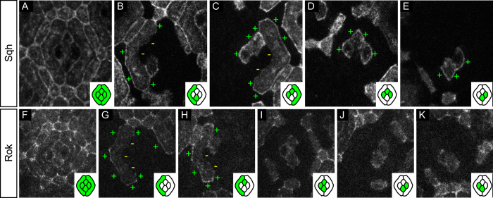
Figure 4. Planar distribution of Sqh and its kinase Rok.
(A–E) Sqh::GFP mosaics. (A) Characteristic distribution of Sqh::GFP in the 32 h APF ommatidial epithelium. (B) Sqh::GFP is enriched on the outer primary pigment cell interfaces (+) and depleted from their inner interfaces (−). (C–E) Outer cone cell interfaces are positive for Sqh::GFP (+). All cone-cone interfaces carry low amounts of Sqh::GFP proteins, preventing us from drawing strong conclusions on the uni- or bilaterality of the protein there. (F–K) Rok::GFP mosaics. (F) Characteristic distribution of Rok::GFP in the 32 h APF ommatidial epithelium. (G,H) Rok::GFP is enriched on the outer primary pigment cell interfaces (+) and depleted from their inner interfaces (−). (H–K) Planar distribution of Rok in cone cells is highly variable, four random samples are presented here.

