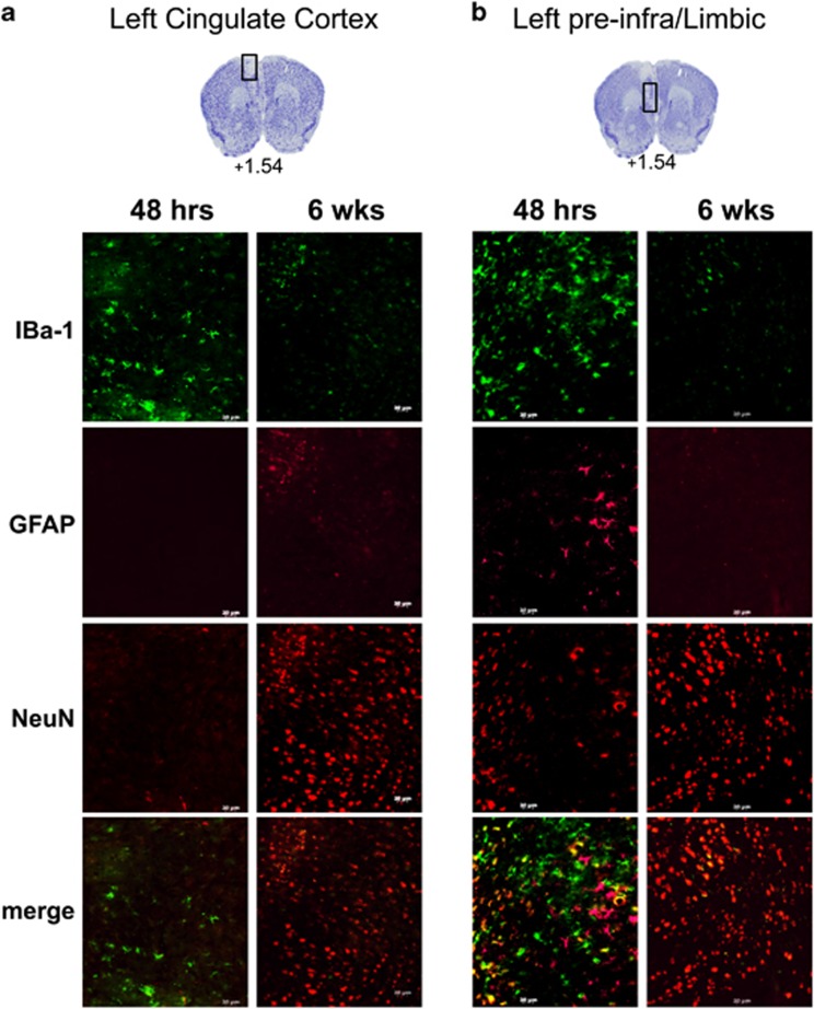
Figure 3.
Cellular changes in the ischemic lesion site over time. Brain sections of the left mPFC (Bregma +1.54) were examined by immunofluorescence at the (a) cingulate gyrus (CG) and (b) pre- and infra-limbic cortex from mice 48 h (left) or 6 weeks (right) following ET-1 microinjection in left mPFC. Representative images of sections stained with anti-IBa1 (microglia), GFAP (astrocyte) and NeuN (neurons). At 48 h post stroke, the lesion site had IBa1-expressing cells in the absence of NeuN-expressing cells (left panels). In contrast at 6 weeks post stroke, the lesion site had NeuN-expressing cells in absence of IBa1 cells (right panels). Scale bar, 20 μm (48 h, n=3; 6 weeks, n=4). ET-1, endothelin-1; mPFC, medial prefrontal cortex.

