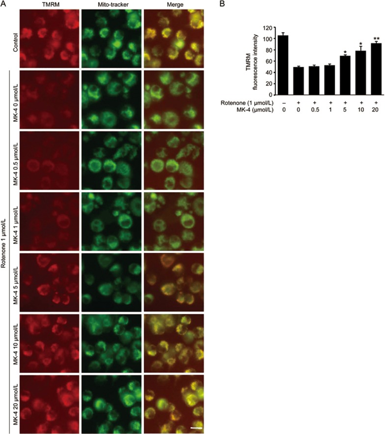
Figure 5.
MK-4 restores the mitochondrial membrane potential damaged by rotenone. (A) Various doses of MK-4 and 1 μmol/L rotenone were administered to BV2 cells as indicated for 24 h. The cells were stained with TMRM and MitoTracker Green. TMRM was used to detect mitochondrial membrane potential. MitoTracker Green was used to detect the mitochondria. The cells were visualized under a fluorescence microscope. Scale bars, 5 μm. (B) The fluorescence intensity of TMRM was analyzed with a multi-detection reader. The data are presented as the mean±SEM from three independent experiments. *P<0.05, **P<0.01 vs the group in which the cells were treated with 1 μmol/L rotenone and 0 μmol/L MK-4, as analyzed by one-way ANOVA.

